When it comes to understanding heart conditions, few terms are as distinct as the 3 sign aortic coarctation.This unique feature plays a crucial role in diagnosing a serious congenital condition known as coarctation of the aorta. At CTS Speciality Hospital, we aim to provide clarity and expert care for patients with this condition. Let’s dive into what the 3 sign aortic coarctation means and why it matters.
What is Coarctation of the Aorta?
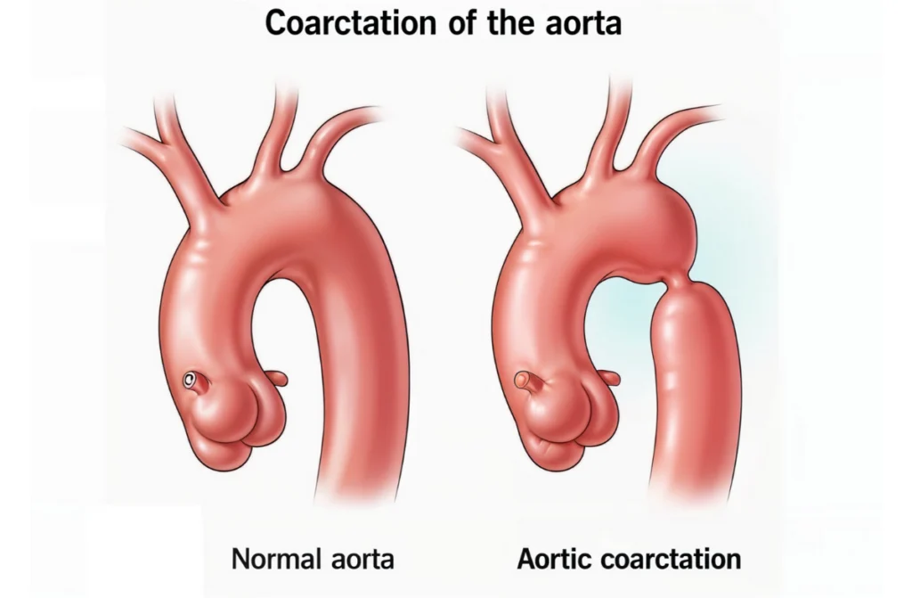
Coarctation of the aorta is a narrowing of the aorta, the large blood vessel responsible for carrying oxygen-rich blood from the heart to the rest of the body. This narrowing can obstruct blood flow, leading to complications. The “3 sign aortic coarctation” is a telltale diagnostic indicator that helps doctors identify this condition on a chest X-ray. This indicator is not just significant but also serves as a cornerstone for early diagnosis and timely intervention.
Coarctation can occur at various points along the aorta but is most commonly found just beyond the arteries that supply blood to the head and arms. This positioning often contributes to the characteristic high blood pressure in the upper body and weaker pulses in the lower body. Recognizing the 3 sign aortic coarctation on imaging is vital for initiating appropriate treatment.
How Common Is Coarctation of the Aorta?
While coarctation of the aorta is not as common as some other congenital heart defects, it’s a significant condition that affects approximately 4 out of every 10,000 live births. Understanding how rare is coarctation of the aorta can help patients and families recognize the importance of specialized care. Early diagnosis, including spotting the 3 sign aortic coarctation on imaging, is essential for successful treatment.
Interestingly, coarctation of the aorta is more prevalent in males than females and is often diagnosed during infancy or early childhood. However, in some cases, the condition may go undetected until adulthood, especially if symptoms are mild or absent during childhood. This highlights the importance of regular health check-ups and advanced diagnostic methods like those available at CTS Speciality Hospital.
What Are the Symptoms of Coarctation of the Aorta?
Symptoms of coarctation of the aorta vary depending on the severity of the narrowing. Common signs include:
- High blood pressure: Especially in the arms, often leading to complications like headaches.
- Weak or absent pulses: Particularly in the legs, indicating restricted blood flow.
- Cold feet or leg pain: Suggestive of reduced oxygen supply to the lower body.
- Headaches or nosebleeds: Caused by elevated blood pressure in the upper body.
These symptoms emphasize how serious is coarctation of the aorta and why prompt medical attention is crucial. In infants, severe coarctation can present as difficulty breathing, poor feeding, or failure to thrive, necessitating immediate medical intervention.
How Serious Is Coarctation of the Aorta?
Understanding how serious is coarctation of the aorta is key to recognizing the urgency of treatment. Left untreated, it can lead to life-threatening complications such as:
- Heart failure: The heart has to work harder to pump blood through the narrowed section.
- Stroke: Elevated blood pressure can increase the risk of stroke, even in young individuals.
- Aneurysm or rupture: Weakening of the aortic wall can lead to potentially fatal complications.
Early intervention can make a significant difference, improving both life expectancy and quality of life. This condition highlights the critical role of the 3 sign aortic coarctation in ensuring timely medical attention.
What Causes Coarctation of the Aorta?
The exact cause of coarctation of the aorta is not fully understood, but it is often linked to congenital factors. Some cases occur alongside other heart defects, such as:
- Bicuspid aortic valve: A common abnormality where the aortic valve has two flaps instead of three.
- Patent ductus arteriosus: A condition where a fetal blood vessel fails to close after birth.
In rare instances, coarctation may develop later in life due to inflammatory conditions or trauma. Identifying the condition through features like the 3 sign aortic coarctation helps pinpoint the problem and determine the best course of action.
How Is Coarctation of the Aorta Diagnosed?
Diagnosis involves a combination of physical examination, imaging, and other tests. The 3 sign aortic coarctation is a critical feature seen on a chest X-ray that suggests the presence of this condition. Additional diagnostic tools include:
- Echocardiogram: To assess heart function and blood flow.
- MRI or CT scans: To get detailed images of the aorta and confirm the extent of the narrowing.
- Cardiac catheterization: To measure blood pressure directly within the heart and arteries.
Recognizing the 3 sign chest x ray coarctation of the aorta ensures accurate diagnosis, paving the way for effective treatment. Regular follow-ups and advanced imaging techniques are crucial for managing this condition over time.
How Rare Is Coarctation of the Aorta?
If you’ve wondered how rare is coarctation of the aorta, it’s considered a rare congenital condition, affecting about 5-8% of people born with congenital heart defects. The rarity makes timely diagnosis, including spotting the 3 sign aortic coarctation, even more vital. Knowing this underscores the importance of expert facilities like CTS Speciality Hospital, where such rare conditions are treated with precision and care.
Patients with undiagnosed coarctation often experience complications as they age, including severe hypertension. Early detection through advanced diagnostic tools ensures that such risks are minimized, even in rare cases.
Tests to Diagnose Coarctation of the Aorta
Doctors use several tests to confirm the diagnosis, including:
- Chest X-ray: Identifies the 3 sign chest x ray coarctation of the aorta by showing the classic ‘3’ shape on the aortic outline.
- Pulse oximetry: Measures oxygen levels in the blood to detect potential discrepancies.
- Electrocardiogram (ECG): Checks for strain or abnormalities in the heart’s electrical activity.
- Doppler ultrasound: Evaluates blood flow patterns and velocity.
Each of these tools plays a role in forming a complete picture of the condition, ensuring precise treatment planning. Recognizing how rare is coarctation of the aorta also emphasizes the need for advanced diagnostic methods.
Surgery to Repair Coarctation of the Aorta
Treatment typically involves surgery or catheter-based interventions to correct the narrowing. Options include:
- Balloon angioplasty: A minimally invasive procedure where a balloon is inflated to widen the narrowed segment.
- Stent placement: A stent is inserted to keep the aorta open permanently.
- Surgical repair: In severe cases, surgeons remove or bypass the narrowed section of the aorta.
These treatments are designed to restore normal blood flow, alleviate symptoms, and prevent long-term complications related to how serious is coarctation of the aorta. Long-term follow-up care is essential to monitor for any recurrence or complications.
The 3 Sign on Chest X-Ray in Coarctation of the Aorta
The 3 sign chest x ray coarctation of the aorta is a distinct pattern formed by:
- Pre-stenotic dilation: The first curve in the ‘3’ represents the area above the narrowing.
- Narrowed segment: The middle of the ‘3’ reflects the actual constriction.
- Post-stenotic dilation: The lower curve signifies the dilation below the narrowing.
This hallmark feature provides critical evidence for diagnosing coarctation of the aorta and planning treatment. Recognizing the 3 sign aortic coarctation ensures prompt medical attention, which is vital given how serious is coarctation of the aorta.
How Does Coarctation Affect the Heart?
Coarctation of the aorta forces the heart to work harder to pump blood through the narrowed vessel. Over time, this can lead to:
- Left ventricular hypertrophy: Thickening of the heart muscle due to increased workload.
- Heart failure: In severe cases, the heart may struggle to pump effectively.
- Aneurysms or rupture: High pressure can weaken the aortic walls, leading to dangerous complications.
The effects on the heart highlight how rare is coarctation of the aorta but also stress its serious nature, emphasizing early intervention.
Conclusion
The 3 sign aortic coarctation is a critical clue in identifying a potentially serious heart condition. Understanding how serious is coarctation of the aorta and how rare is coarctation of the aorta underscores the importance of timely diagnosis and treatment. At CTS Speciality Hospital, we are committed to providing exceptional care, from diagnosis to recovery.